Overview
what it is and why it matters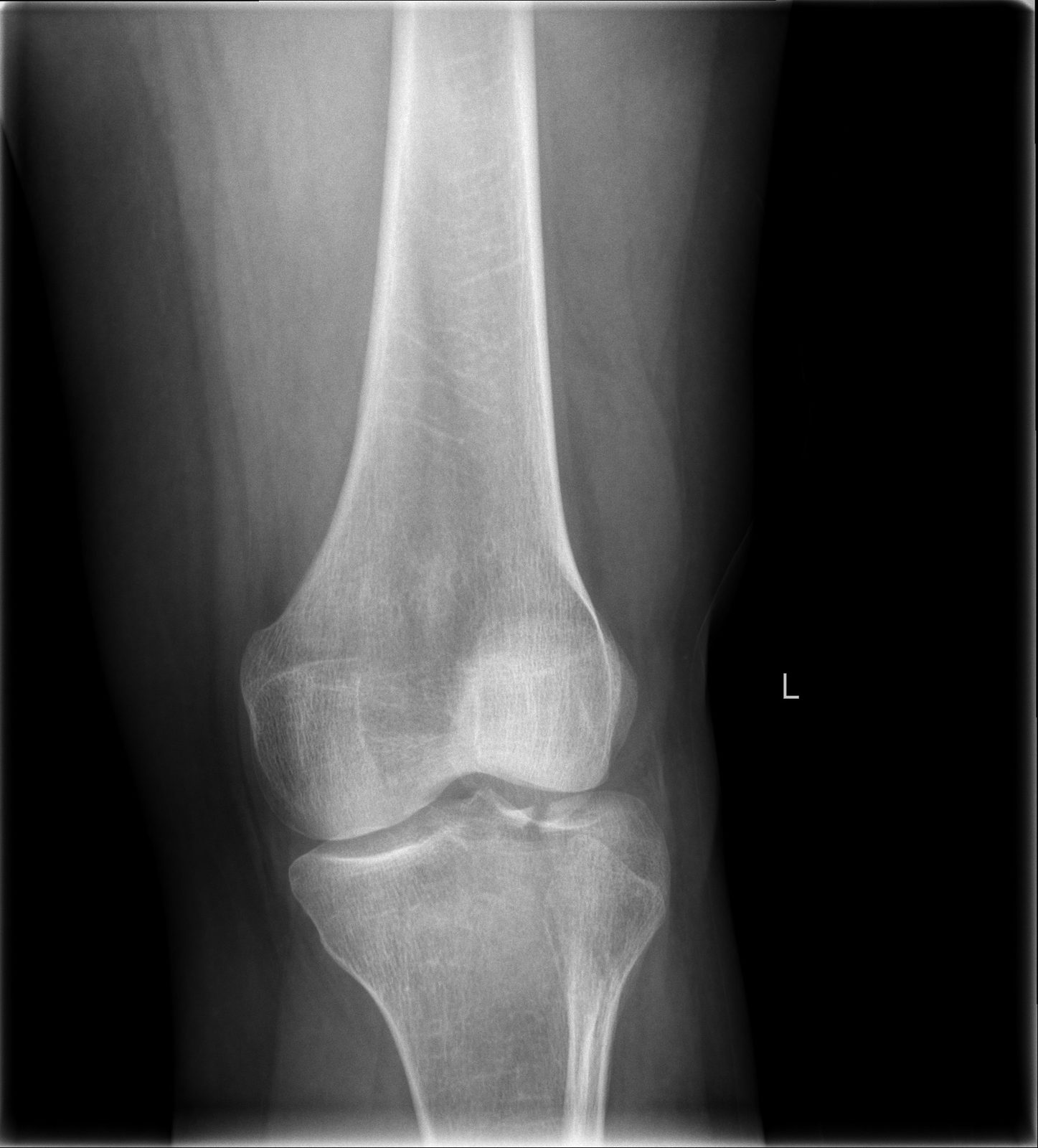
The tibial plateau is the flat upper surface of the tibia that articulates with the femur and bears body weight across the knee joint. Fractures here occur from axial load combined with valgus or varus force — classically a pedestrian struck by a car bumper ("bumper fracture"), a fall from height, or a sports collision. The Schatzker classification (types I–VI) describes fracture patterns from simple lateral split to severe bicondylar crush.
Associated injuries — meniscal tears, ligament disruptions, and compartment syndrome — are common and must be systematically evaluated.
Diagnosis
exam first, imaging secondKnee swelling (hemarthrosis), inability to bear weight, and tenderness over the proximal tibia. CT scan with 3D reconstruction is mandatory for surgical planning. MRI evaluates the menisci and ligaments. Vascular examination of the limb is critical — popliteal artery injury must be excluded.
Treatment Path
how care progresses at OSI1
Non-operative management
For minimally displaced fractures (depression < 3–5 mm, acceptable alignment) in older, lower-demand patients: non-weight-bearing for 8–12 weeks with close radiographic follow-up.
Surgical Options at OSI
if non-operative care isn't enoughSignificant articular depression, condylar widening, combined ligamentous instability, or osteomyelitis and septic…">open fractures require surgical intervention.
Providers Who Treat Tibial Plateau Fracture
sports-medicine team

David B. Templin, M.D.

Trent Twitero, M.D.
Further Reading
authoritative sourcesExternal patient-education references and related OSI pages for additional background: