Overview
The anterior cruciate ligament, or ACL, is one of the four major stabilizing ligaments of the knee. It runs diagonally inside the joint, connecting the femur (the thigh bone) to the tibia (the shin bone), and its job is to keep the tibia from sliding forward on the femur — especially during the pivot, cut, and landing motions that dominate athletics. A tear of the ACL almost always happens without contact: a sudden deceleration, a change of direction, or a landing where the knee collapses inward. Patients typically describe an audible pop, rapid swelling within hours, and a knee that can no longer be trusted to support them.
Women are several times more likely than men to tear the ACL, the difference explained by landing mechanics, neuromuscular patterns, and the shape of the intercondylar notch through which the ligament runs. Once the ligament is torn it does not heal on its own, because it lives inside the synovial fluid of the joint and lacks the blood supply needed to knit itself back together. How that matters depends on the demands the patient puts on the knee: pivoting sports usually require reconstruction, while lower-demand daily life can sometimes be managed without surgery.
Diagnosis & Evaluation
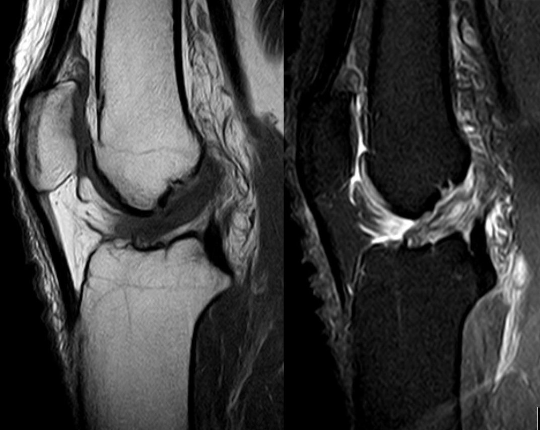
The Lachman test is the most sensitive examination maneuver — the examiner feels for increased anterior tibial translation compared to the other side. The anterior drawer and pivot shift tests provide additional information. MRI confirms the diagnosis and evaluates for associated meniscal, cartilage, and collateral ligament injuries, which occur in the majority of ACL tears.
Non-Surgical Treatment
Bracing & acute management
Knee brace for comfort, ice, elevation, and protected weight-bearing in the acute phase.
Physical therapy
Quadriceps and hamstring rehabilitation restores strength and may be sufficient for lower-demand, older patients who are willing to modify activity and accept some instability.
Activity modification
Some patients with ACL-deficient knees can return to straight-line activities but must avoid pivoting sports.
When Surgery Is Considered
ACL reconstruction is recommended for active patients who wish to return to pivoting or cutting sports, young patients with a long active life ahead, and patients with associated injuries (meniscal tear, cartilage damage) that require surgical addressing.
ACL reconstruction
The torn ligament is replaced with a tendon graft — most commonly the patellar tendon (bone-tendon-bone), hamstring tendons, or quadriceps tendon. The graft is fixed in tunnels drilled in the femur and tibia and becomes a functional ligament through dedicated rehabilitation.
If non-operative care is not enough, these procedures are offered by the OSI team for this condition:
Providers Who Treat Acl Tear


David B. Templin, M.D.

Trent Twitero, M.D.
Further Reading
authoritative sourcesExternal patient-education references and related OSI pages for additional background: