Overview
what it is and why it matters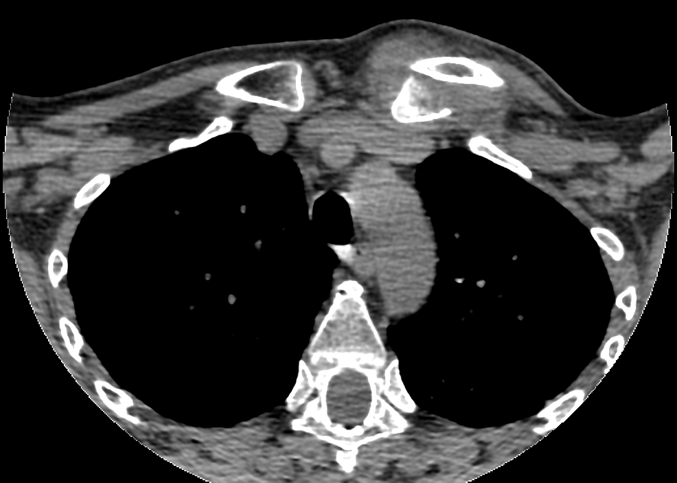
The sternoclavicular (SC) joint is where the medial end of the clavicle articulates with the sternum (breastbone). SC injuries range from mild sprains (most common) to complete dislocations, which can be anterior (less dangerous) or posterior (serious — posterior dislocation can compress the trachea, esophagus, or major vessels). SC dislocations are uncommon and typically result from high-energy trauma.
Diagnosis
exam first, imaging secondMedial clavicular pain, swelling, and prominence. Plain X-rays are technically difficult to interpret; CT scan is the definitive study for suspected dislocation. For posterior dislocations, a thoracic surgery consult may be needed given proximity to great vessels.
Treatment Path
how care progresses at OSI1
Sling and symptomatic care
Appropriate for sprains and anterior dislocations — most are managed non-operatively with gradual return to activity.
2
Closed reduction
Attempted under procedural sedation or general anesthesia for acute dislocations with a cardiothoracic surgeon available for posterior cases.
Surgical Options at OSI
if non-operative care isn't enoughPosterior SC dislocations causing airway or vascular compromise require emergency surgery. Chronic SC instability causing pain and functional limitation is addressed with reconstruction.
Providers Who Treat Sternoclavicular Injury
sports-medicine team

David B. Templin, M.D.

Trent Twitero, M.D.
Further Reading
authoritative sourcesExternal patient-education references and related OSI pages for additional background: