Overview
what it is and why it matters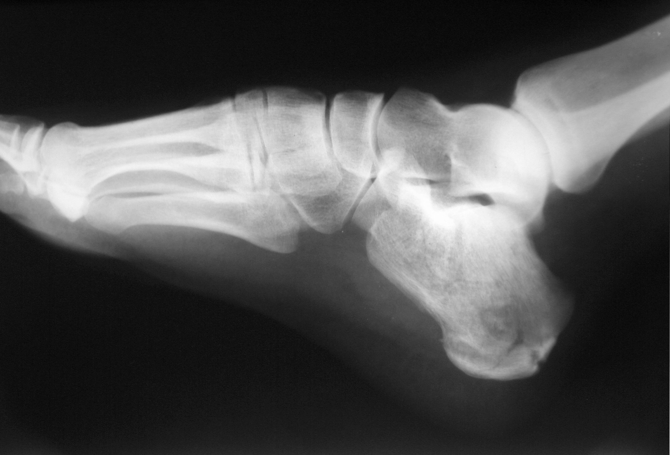
Calcaneus fractures are the most common tarsal bone fractures and occur from axial loading — typically a fall from height. Bilateral fractures occur in 10% of cases (and should prompt evaluation for associated lumbar spine fracture). Most calcaneus fractures are intra-articular, involving the posterior facet of the subtalar joint. The Bohler angle (normally 20–40°) collapses in displaced fractures. Severe soft-tissue injury and fracture blisters are common.
Diagnosis
exam first, imaging secondLateral calcaneal X-ray and axial (Harris) view. CT is essential for surgical planning, characterizing joint involvement (Sanders classification), fracture pattern, and degree of comminution. A full lumbar spine X-ray should be obtained for high-energy falls.
Treatment Path
how care progresses at OSINon-weight-bearing and elevation
Initial management for all calcaneus fractures to control swelling.
Functional rehabilitation
For non-displaced or extra-articular fractures: early range-of-motion with NWB for 6–8 weeks.
Definitive non-operative management
Extra-articular fractures and some intra-articular fractures in elderly or diabetic patients with poor bone quality are managed non-operatively.
Surgical Options at OSI
if non-operative care isn't enoughDisplaced intra-articular fractures (Sanders II-IV) in physiologically young, healthy patients with good bone quality typically benefit from ORIF to restore subtalar joint congruity.
Further Reading
authoritative sourcesExternal patient-education references and related OSI pages for additional background: