Overview
what it is and why it matters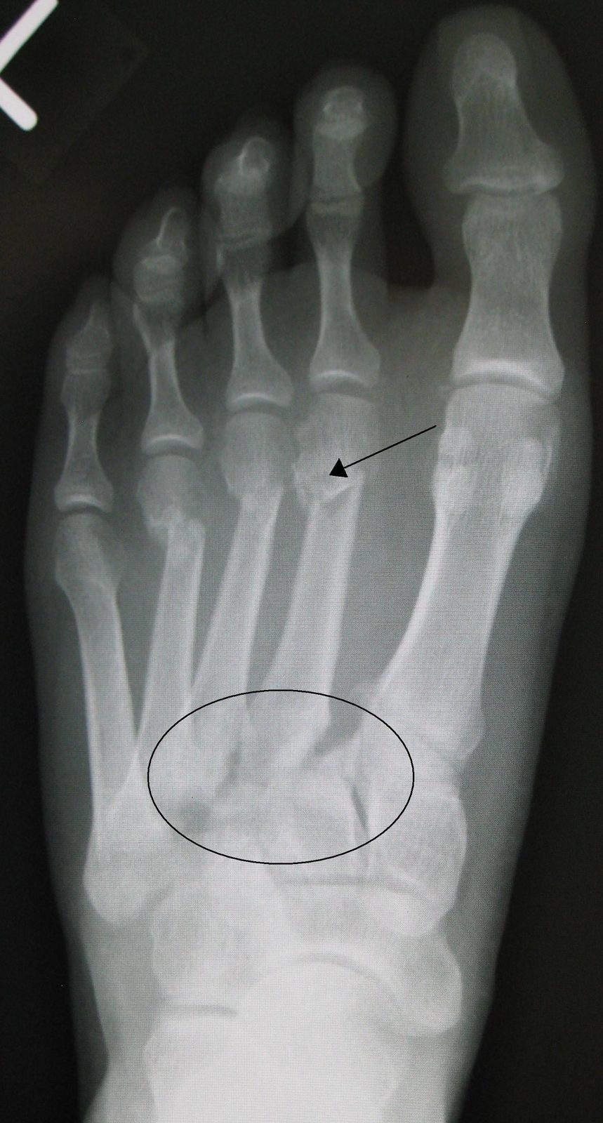
The Lisfranc joint is the tarsometatarsal (TMT) articulation that anchors the midfoot. Lisfranc injuries — ranging from subtle ligamentous sprains to dramatic fracture-dislocations — are commonly missed, and even a "subtle" Lisfranc ligament disruption leads to progressive midfoot arthritis and disability if not recognized and treated. The injury occurs from axial loading with the foot in plantarflexion (car pedal impact) or from a seemingly minor twist (stepping off a curb).
The classic subtle presentation is a midfoot sprain that fails to improve. Plantar ecchymosis over the arch is a pathognomonic sign.
Diagnosis
exam first, imaging secondNon-weight-bearing X-rays are frequently normal. Weight-bearing AP foot X-rays are essential — diastasis between the first and second metatarsal bases (>2 mm) confirms the diagnosis. CT defines bony detail. MRI is most sensitive for purely ligamentous injuries. Plantar arch bruising on exam is highly suspicious.
Treatment Path
how care progresses at OSINon-weight-bearing cast
Purely ligamentous injuries in low-demand patients without diastasis may be managed 6–10 weeks NWB with close radiographic follow-up.
Surgical Options at OSI
if non-operative care isn't enoughAny diastasis, fracture-dislocation, or ligamentous injury with demonstrated instability in an active patient requires surgical stabilization.
Further Reading
authoritative sourcesExternal patient-education references and related OSI pages for additional background: