Overview
what it is and why it matters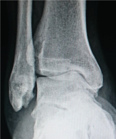
Ankle osteoarthritis is far less common than hip or knee OA and is usually post-traumatic — the result of prior fracture, recurrent instability, or osteochondral lesions rather than primary wear. It is a major cause of long-term disability after ankle fractures. The tibiotalar joint degenerates with cartilage loss, subchondral sclerosis, and osteophyte formation, causing pain, stiffness, and difficulty walking on uneven surfaces.
Diagnosis
exam first, imaging secondWeight-bearing AP, lateral, and mortise ankle X-rays are the primary imaging study. CT provides superior detail of osteophytes and subchondral cysts. MRI assesses cartilage and soft-tissue pathology. Alignment of the hindfoot must be assessed — varus or valgus deformity drives asymmetric ankle loading.
Treatment Path
how care progresses at OSIActivity modification
Avoiding prolonged weight-bearing, impact sports.
Supportive footwear / orthosis
Rocker-bottom sole reduces dorsiflexion demand and ankle motion. AFO bracing for severe cases.
Physical therapy
Gait retraining, calf stretching.
Ankle corticosteroid injection
Provides temporary relief; useful for flares.
Viscosupplementation (hyaluronic acid)
Limited evidence but may provide months of relief.
Surgical Options at OSI
if non-operative care isn't enoughSurgery is offered when pain significantly limits function and conservative measures have failed.
Further Reading
authoritative sourcesExternal patient-education references and related OSI pages for additional background: