Overview
what it is and why it matters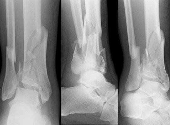
A pilon fracture is a high-energy axial compression fracture of the distal tibia that shatters the tibial plafond (articular surface). It occurs from falls from height or ski injuries, and is associated with significant soft-tissue injury. Pilon fractures represent one of the most challenging fractures to treat in orthopedic surgery — articular comminution, bone loss, and soft-tissue compromise must all be addressed. Staged management (temporary external fixation until soft-tissue swelling resolves, then definitive ORIF) is standard.
Diagnosis
exam first, imaging secondAP, lateral, and mortise ankle X-rays. CT with 3D reconstructions is mandatory for surgical planning, defining articular fragments, degree of comminution, and impaction. Soft-tissue assessment (fracture blisters, skin viability) is critical before surgical timing is determined.
Treatment Path
how care progresses at OSIExternal fixation (spanning)
Initial temporizing management to restore length and alignment while soft tissues recover. Not definitive.
Surgical Options at OSI
if non-operative care isn't enoughVirtually all displaced pilon fractures require staged surgical reconstruction once the soft-tissue envelope permits.
Further Reading
authoritative sourcesExternal patient-education references and related OSI pages for additional background: