Overview
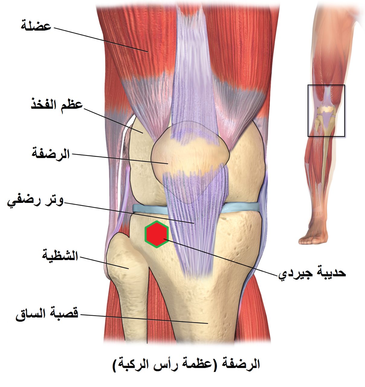
The tibial plateau is the top surface of the tibia that forms the lower half of the knee joint. Plateau fractures often result from a valgus- or varus-directed blow, commonly in pedestrian-versus-car injuries or falls. Joint-surface depression and split patterns are typical.
Surgery is indicated for significant articular depression, split fractures that affect the joint, and bicondylar injuries. Restoration of a smooth, level joint surface is the primary goal because step-offs and residual depression drive post-traumatic arthritis.
Why it's done
Tibial plateau ORIF is typically considered when imaging and the clinical picture together indicate that the fracture will not reliably heal or function without surgical stabilization. Common indications include:
Depressed articular fragment
A plateau step-off accelerates cartilage wear.
Displaced split fracture
Widens the plateau and destabilizes the knee.
Bicondylar fracture
Involves both plateaus and is inherently unstable.
Soft-tissue compromise or compartment syndrome
May require staged external fixation first.
Open fracture
Urgent debridement and fixation.
How it works
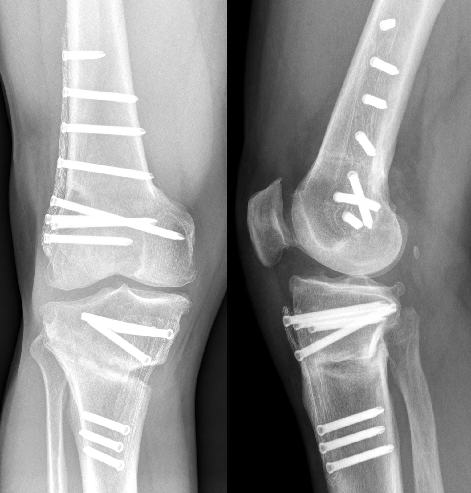
For simple split fractures, a small incision allows the fragment to be lifted back to its original position and secured with screws and a buttress plate.
For fractures with central joint depression, a cortical window below the joint is used to elevate the depressed cartilage with a bone tamp. The resulting defect is filled with bone graft or bone-graft substitute, and the construct is stabilized with a locking plate. In bicondylar injuries, dual medial and lateral plates may be used.
Recovery
Protected weight-bearing is typical for ten to twelve weeks. Knee range-of-motion exercises begin early, usually within the first week, because stiffness is a major concern. Physical therapy is important throughout recovery. Union is typically seen by three months. Post-traumatic arthritis is a known long-term risk; total knee replacement may become an option later if symptoms develop. Hardware is left unless it becomes symptomatic.
Contact
For questions about this procedure or to schedule an evaluation, call the office at (830) 625-0009 or request an appointment online.
Further Reading
External patient-education references and related OSI pages for additional background: