Overview
what it is and why it matters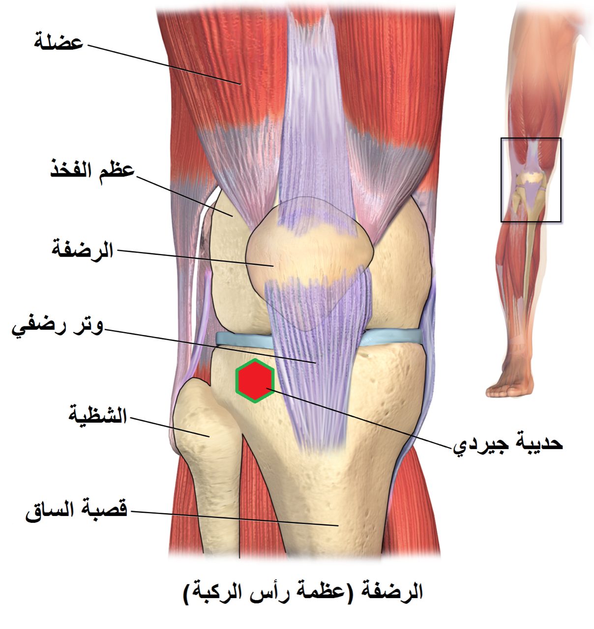
Patellar instability describes a spectrum from subtle subluxation (partial slip) to complete dislocation of the patella from the trochlear groove. First-time dislocations typically occur in adolescents and young adults during a pivoting movement or direct blow. A dislocation tears the medial patellofemoral ligament (MPFL) — the primary soft-tissue restraint preventing lateral patellar displacement — and damages the medial facet of the patella and the lateral trochlear wall.
Recurrence rates after a first dislocation are 15–40% in adults and higher in adolescents. Risk factors for recurrence include trochlear dysplasia, high patella (patella alta), and increased TT-TG distance.
Diagnosis
exam first, imaging secondAcute dislocation presents with lateral displacement of the patella (usually reduces spontaneously), hemarthrosis, and medial retinacular tenderness. The patellar apprehension test (lateral pressure on the patella with knee extended) is positive. X-rays include a merchant/sunrise view and lateral view. MRI evaluates MPFL integrity, osteochondral injury, and trochlear anatomy.
Treatment Path
how care progresses at OSI1
Reduction (if still dislocated)
Gentle extension of the knee with medial pressure on the patella to relocate it — performed in the emergency setting.
2
Bracing
Lateral restraint brace for 4–6 weeks after acute dislocation.
3
Physical therapy
VMO and hip abductor strengthening is the cornerstone of non-operative management after a first dislocation.
Surgical Options at OSI
if non-operative care isn't enoughFirst-time dislocation with a significant osteochondral fracture requires early surgery. Recurrent instability — especially with anatomic risk factors — is the primary indication for reconstruction.
Primary procedure
MPFL reconstruction
The torn medial patellofemoral ligament is reconstructed using a gracilis or quadriceps tendon graft — the most commonly performed procedure for recurrent instability.
Learn about this procedure →Additional option
Tibial tubercle osteotomy
Repositioning the patellar tendon attachment to correct tracking.
Learn about this procedure →Providers Who Treat Patellar Instability & Dislocation
sports-medicine team

David B. Templin, M.D.

Trent Twitero, M.D.
Further Reading
authoritative sourcesExternal patient-education references and related OSI pages for additional background: