Overview
what it is and why it matters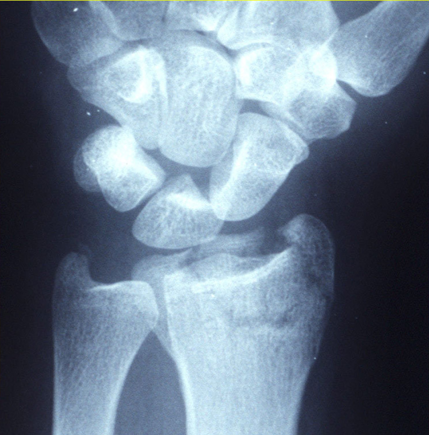
A distal radius fracture is a break of the radius — the thicker of the two forearm bones — just above the wrist. It is the most common fracture seen in orthopedic practice, and the mechanism is almost always the same: a fall in which the person instinctively puts out a hand to catch themselves, driving the wrist backward and concentrating the force at the weakest point of the bone. When the broken end tips backward, the wrist takes on a characteristic bent-back profile known as a Colles’ fracture; the less common Smith’s fracture tips the opposite way.
What looks like a single injury is actually a spectrum. A minimally displaced break in an older patient with thinning bone behaves very differently from a shattered intra-articular fracture in a younger patient after a fall from height. Treatment depends on where the bone has broken, how far out of position it sits, whether the joint surface has been disrupted, and the demands the patient places on the wrist — which is why two people with similar-looking X-rays may be offered different plans.
Diagnosis
exam first, imaging secondPA and lateral wrist X-rays diagnose the fracture and characterize displacement, angulation (radial inclination, volar tilt), shortening, and articular involvement. CT is obtained for complex intra-articular patterns to plan fixation. Associated TFCC or scapholunate injuries are considered whenever there is ulnar-sided pain or instability.
Treatment Path
how care progresses at OSI1
Closed reduction and cast immobilization
Acceptable alignment in low-demand patients (radial shortening <3 mm, volar tilt within 5° of neutral, no intra-articular step-off >2 mm) can be managed in a short-arm cast for 4–6 weeks.
Surgical Options at OSI
if non-operative care isn't enoughSurgery is indicated for unacceptable alignment, unstable fractures that re-displace in cast, intra-articular step-off >2 mm, or in active patients who need reliable healing in anatomic position.
Providers Who Treat Distal Radius Fracture
sports-medicine team

David B. Templin, M.D.

Trent Twitero, M.D.
Further Reading
authoritative sourcesExternal patient-education references and related OSI pages for additional background: