Overview
what it is and why it matters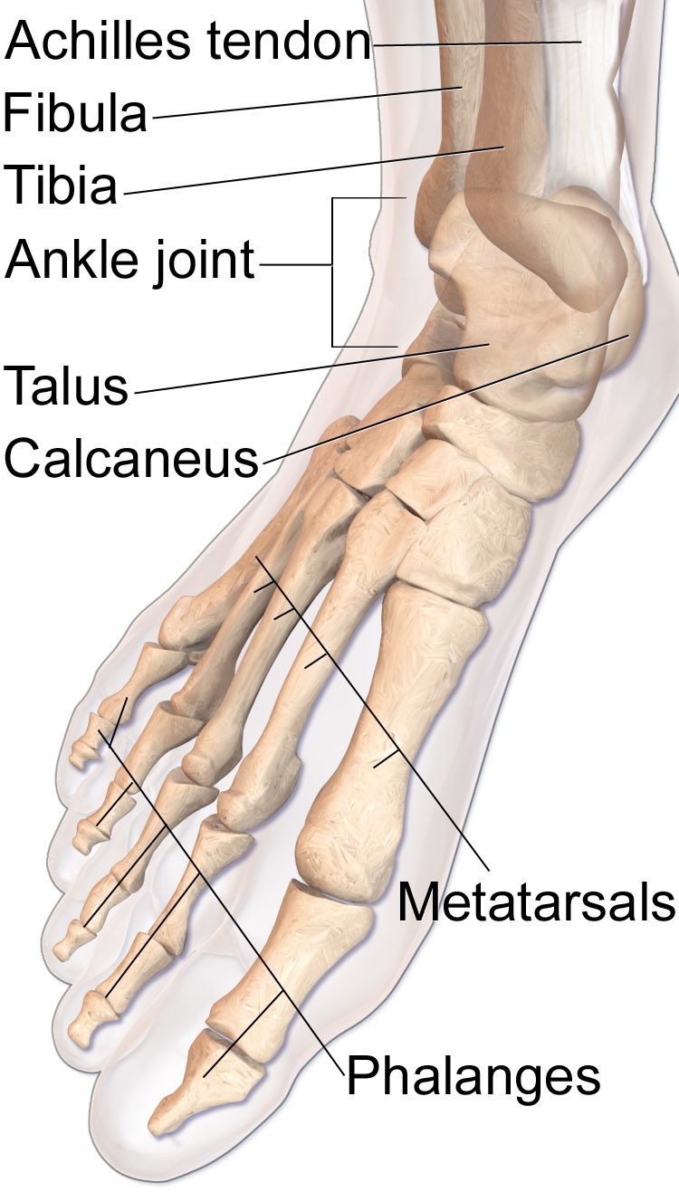
The peroneal tendons (brevis and longus) run in a groove behind the lateral malleolus and evert the foot, protecting against ankle inversion. Longitudinal splits of the peroneus brevis — especially at the retromalleolar groove — are the most common peroneal tendon injury. They are often associated with lateral ankle instability and can be subtle, presenting as persistent lateral ankle pain after an "ankle sprain" that fails to resolve.
Diagnosis
exam first, imaging secondTenderness posterior and inferior to the lateral malleolus, pain with resisted eversion, and peroneal squeeze test. MRI is the definitive test, showing longitudinal split tears ("flattened" or bifid peroneus brevis on axial cuts). Dynamic ultrasound can demonstrate subluxation of the tendons over the malleolus.
Treatment Path
how care progresses at OSIImmobilization
4–6 weeks of casting or walking boot for partial tears.
Physical therapy
Eversion strengthening and proprioceptive retraining.
Surgical Options at OSI
if non-operative care isn't enoughFull-thickness longitudinal tears, subluxing tendons, and failed conservative care require surgical management.
Further Reading
authoritative sourcesExternal patient-education references and related OSI pages for additional background: