Overview
what it is and why it matters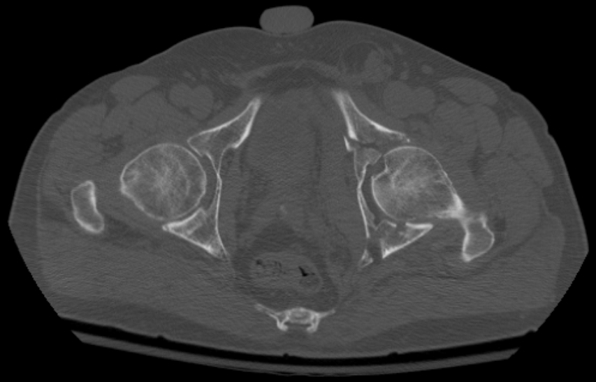
The acetabulum (hip socket) is the cup-shaped portion of the pelvis that receives the femoral head. Acetabular fractures are typically caused by high-energy mechanisms — falls from height or high-impact sports collisions — in which force is transmitted up the femoral shaft into the socket. The Letournel and Judet classification system describes 10 fracture types based on which walls and columns of the acetabulum are involved.
Because the acetabulum is the weight-bearing surface of the hip joint, accurate reconstruction is essential to prevent post-traumatic arthritis.
Diagnosis
exam first, imaging secondHip pain, inability to bear weight, and potential hip dislocation (the femoral head may be displaced from the socket). Plain X-rays of the pelvis — AP, obturator oblique, and iliac oblique (Judet views) — characterize most fractures. CT scan is essential for operative planning and evaluating articular comminution.
Treatment Path
how care progresses at OSI1
Non-operative management
Reserved for non-displaced fractures, fractures with a stable hip joint on examination, and patients who are not surgical candidates. Protected weight-bearing for 8–12 weeks.
Surgical Options at OSI
if non-operative care isn't enoughDisplaced fractures with joint incongruity, hip instability, or intra-articular fragments require surgical reconstruction. The goal is restoring the articular surface to within 2 mm of anatomic position to minimize arthritis risk.
Providers Who Treat Acetabular Fracture
sports-medicine team

David B. Templin, M.D.

Trent Twitero, M.D.
Further Reading
authoritative sourcesExternal patient-education references and related OSI pages for additional background: