Overview
Distal femur fractures occur just above the knee. They commonly follow high-energy trauma in younger patients and low-energy falls in older, osteoporotic patients. Many patterns cross into the knee joint surface.
Non-operative treatment rarely holds alignment in these fractures. Surgery allows the limb to be brought back to length and rotation, the joint surface to be reconstructed, and early motion started, which is especially important for knee function.
Why it's done
Distal femur ORIF is typically considered when imaging and the clinical picture together indicate that the fracture will not reliably heal or function without surgical stabilization. Common indications include:
Displaced or comminuted fracture
Closed methods cannot reliably maintain alignment.
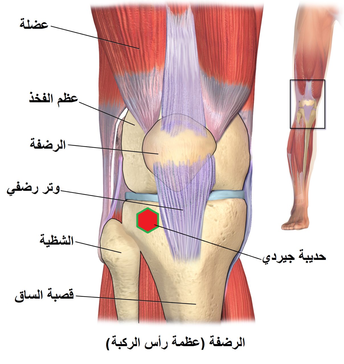
Intra-articular extension
Joint surface step-off accelerates knee arthritis.
Periprosthetic fracture above a knee replacement
Special fixation techniques are required to protect the implant.
Open fracture
Open injuries require urgent washout and stabilization.
Inability to tolerate prolonged non-weight-bearing
Surgery allows earlier mobilization.
How it works
For extra-articular or simple intra-articular patterns, a retrograde intramedullary nail can be passed through a small incision at the front of the knee. For complex intra-articular fractures, a pre-contoured lateral locking plate is applied through a longer incision on the outside of the thigh.
The joint surface is reconstructed first with lag screws, and then the reconstructed block is connected to the femoral shaft with the plate or nail. Fluoroscopy confirms length, rotation, and alignment.
Recovery
Most patients are kept toe-touch or partial weight-bearing for six to twelve weeks depending on the pattern. Knee motion begins early with a continuous passive motion machine or formal therapy to prevent stiffness. Union is typically seen by three to four months. Periprosthetic fractures and highly comminuted patterns may take longer. Hardware is rarely removed.
Contact
For questions about this procedure or to schedule an evaluation, call the office at (830) 625-0009 or request an appointment online.
Further Reading
External patient-education references and related OSI pages for additional background: