Overview
The tibia is the main weight-bearing bone of the lower leg. Shaft fractures often result from high-energy mechanisms such as falls or sports injuries. Because the tibia lies just under the skin along its front surface, open fractures are common.
Intramedullary nailing is the workhorse treatment for displaced tibial shaft fractures. It restores length and alignment through small incisions without directly exposing the fracture, which preserves the healing biology.
Why it's done
Tibial shaft intramedullary nail is typically considered when imaging and the clinical picture together indicate that the fracture will not reliably heal or function without surgical stabilization. Common indications include:
Displaced or unstable shaft fracture
Alignment cannot be reliably held in a cast.
Open fracture
Urgent irrigation, debridement, and stabilization.
Segmental fracture pattern
Two fracture levels are inherently unstable.
Polytrauma
Rapid stabilization aids mobilization and overall care.
Nonunion from prior non-operative care
Nailing plus bone grafting salvages most cases.
How it works
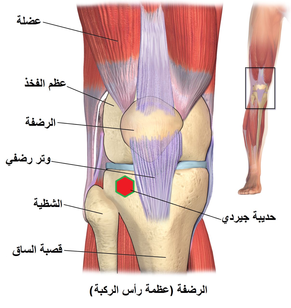
Through a small incision at the front of the knee (either patellar tendon-splitting, medial parapatellar, or suprapatellar), a guidewire is passed down the medullary canal of the tibia under fluoroscopic guidance. The canal is progressively reamed to accept a solid or cannulated nail.
Interlocking screws above and below the fracture control rotation and length. Associated fibular fractures rarely need fixation. Fluoroscopy confirms final alignment, rotation, and nail position.
Recovery
Most isolated tibial shaft fractures allow weight-bearing as tolerated shortly after surgery, particularly with modern locked nails. Early knee and ankle motion begins immediately. Union is typically seen by three to six months. Anterior knee pain after tibial nailing is common and sometimes requires nail removal after healing. Delayed union and malrotation are known complications.
Contact
For questions about this procedure or to schedule an evaluation, call the office at (830) 625-0009 or request an appointment online.
Further Reading
External patient-education references and related OSI pages for additional background: