Overview
what it is and why it matters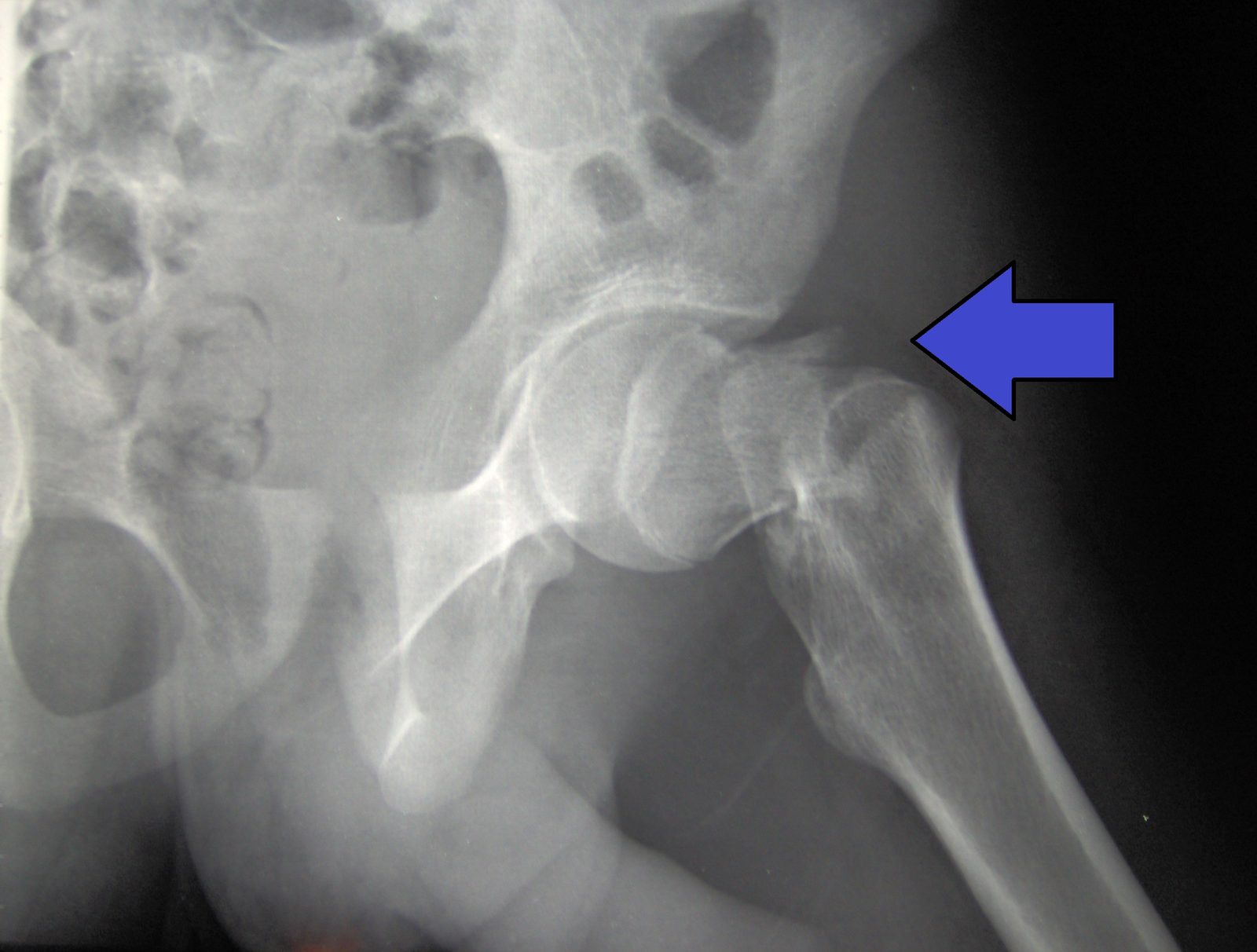
Intertrochanteric fractures occur between the two bony prominences (trochanters) at the upper femur, just below the femoral neck. Like femoral neck fractures, they are most common in elderly women with osteoporosis after a ground-level fall. They are classified as stable or unstable based on whether the fracture pattern allows the fragments to bear load after fixation.
Unlike femoral neck fractures, the blood supply to the femoral head is rarely threatened by intertrochanteric fractures, so fixation — rather than replacement — is the most common surgical approach.
Diagnosis
exam first, imaging secondPresentation mirrors femoral neck fracture: inability to bear weight, shortened and externally rotated extremity, proximal thigh pain. AP pelvis and lateral hip X-rays are the primary imaging studies. CT is used for comminuted or complex patterns to plan fixation strategy.
Treatment Path
how care progresses at OSI1
Non-operative management
Appropriate only for patients who cannot tolerate any anesthetic or surgical procedure, given the high complication rate of prolonged bed rest in elderly patients.
Surgical Options at OSI
if non-operative care isn't enoughSurgical fixation is the standard for virtually all intertrochanteric fractures in patients who can tolerate surgery. Early fixation (within 24–48 hours) improves outcomes.
Providers Who Treat Intertrochanteric Fracture
sports-medicine team

David B. Templin, M.D.

Trent Twitero, M.D.
Further Reading
authoritative sourcesExternal patient-education references and related OSI pages for additional background: