Overview
what it is and why it matters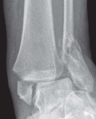
The ankle is a ring — a mortise formed by the lower ends of the tibia and fibula cradling the talus, the small bone of the hindfoot that sits between them. Like any ring, the ankle can tolerate a single break and still hold its shape, but once it is broken in two places the structure loses its ability to keep the talus centered, and the joint surfaces begin to slide out of alignment under load. Ankle fractures typically result from a twisting mechanism — rolling the foot inward or outward during a misstep off a curb or an athletic pivot — and the direction the foot twisted at the moment of injury dictates exactly where the bone breaks.
The decisive question in any ankle fracture is not simply whether a bone is broken, but whether the ring is stable: can the ankle hold the talus in position while it heals, or will the joint drift under body weight? Stability drives the decision between a boot and surgery far more than how dramatic the X-ray looks. A single break that leaves the mortise intact usually heals in a boot; a break that disrupts the ring in two places almost always needs to be fixed in the operating room to restore alignment.
Diagnosis
exam first, imaging secondOttawa Ankle Rules guide X-ray ordering. AP, lateral, and mortise views characterize the fracture pattern. CT is added for complex pilon fractures, posterior malleolus involvement, or preoperative planning. Gravity stress view or external rotation stress X-ray assesses mortise stability in isolated fibula fractures.
Treatment Path
how care progresses at OSIShort-leg cast / walking boot
Stable isolated lateral malleolus fractures without mortise widening can be managed non-operatively with a removable boot and early weight-bearing.
Surgical Options at OSI
if non-operative care isn't enoughAny mortise instability, bimalleolar equivalents, and fractures with talar shift require surgical fixation to restore joint congruity.
Further Reading
authoritative sourcesExternal patient-education references and related OSI pages for additional background: