Overview
what it is and why it matters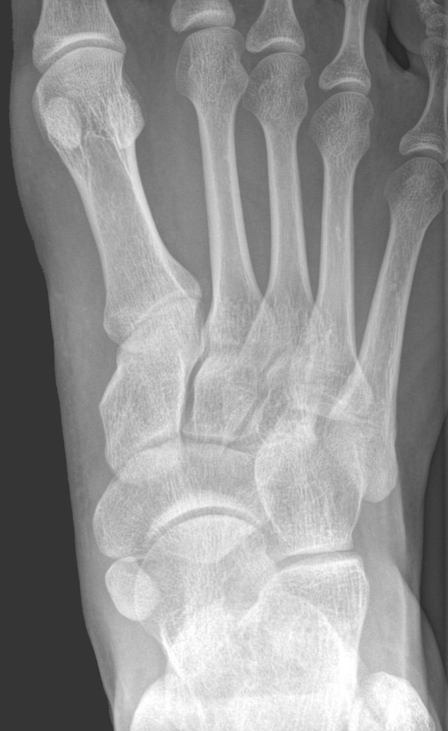
Navicular stress fractures are the highest-risk stress fractures in athletes. The central third of the navicular is avascular ("watershed zone") and slow to heal. They occur predominantly in sprinting and jumping athletes (basketball, track and field) and typically present as vague midfoot pain that worsens with activity. They are frequently missed on initial plain X-rays.
Delayed diagnosis and inadequate treatment result in complete fracture, displacement, and nonunion with long-term disability.
Diagnosis
exam first, imaging secondPoint tenderness at the "N-spot" (dorsal navicular). X-rays are insensitive. MRI is the gold standard for early diagnosis. CT confirms fracture pattern, displacement, and informs surgical planning. Any athlete with midfoot pain and a risk profile for navicular stress fracture should have MRI before returning to activity.
Treatment Path
how care progresses at OSIStrict non-weight-bearing cast
6–8 weeks NWB in a short-leg cast is the minimum for incomplete (type I) and many complete undisplaced (type II) fractures. This is mandatory, not optional — premature weight-bearing causes nonunion.
Surgical Options at OSI
if non-operative care isn't enoughDisplaced fractures (type III), nonunions, and high-performance athletes who need reliable rapid healing opt for surgical fixation.
Further Reading
authoritative sourcesExternal patient-education references and related OSI pages for additional background: